
|
|
|
Diagnosis This is an extremely rare, and little known clinical and histological subtype of Paget's disease of the nipple. Upon first inspection, a diagnosis of melanoma in situ is entertained. Yet the location should clue the pathologist in to the possibility of Paget's disease. Indeed, the immunoperoxidase studies are indispensible in this case as is the negative control. A problem with this case is the background interference of the melanin pigment. One solution is to bleach the slides and then apply the immnoperoxidase stains. Another possibility, performed in this case, is to use a red chromogen with the antibodies. Examination of the negative control is very important in this case since the melanin pigment is so prominent on all slides. An S100 immunoperoxidase stain was negative within the The differential diagnosis includes melanoma, pigmented Bowen's disease, and less likely pemphigus vulgaris with secondary pigment incontinence. References Am J Dermatopathol 2002;245:189-198. For additional case studies in general surgical pathology and clinical pathology, please visit our sister website, GotPath?
|
Last Updated January 26, 2005
Send Emails to
Webmaster at DermpathMD
Read the Medical Disclaimer
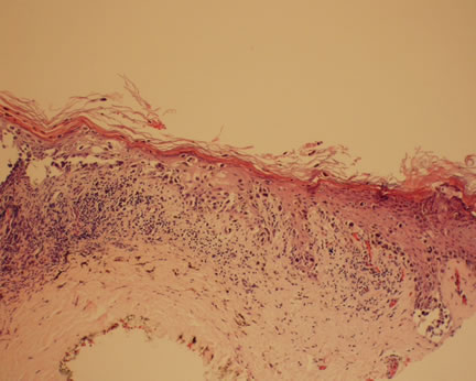
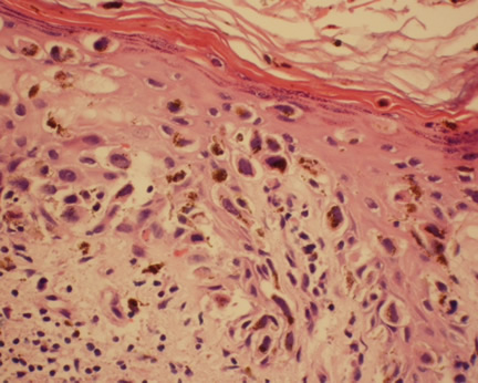
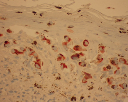 Image 3 EMA decorates many of the atypical intraepithelial cells.